Introduction
Uterine fibroids, medically known as leiomyomas, are common benign tumors that develop in the muscular wall of the uterus. Over time, some fibroids undergo degeneration and become hardened through calcium deposition, resulting in what is referred to as calcified uterine leiomyoma. While these calcified growths are often considered less active than their non-calcified counterparts, hormonal influence still plays a significant role in their development, progression, and clinical presentation. Understanding how hormones affect calcified uterine leiomyoma is essential for both patients and healthcare providers when considering management and treatment options.
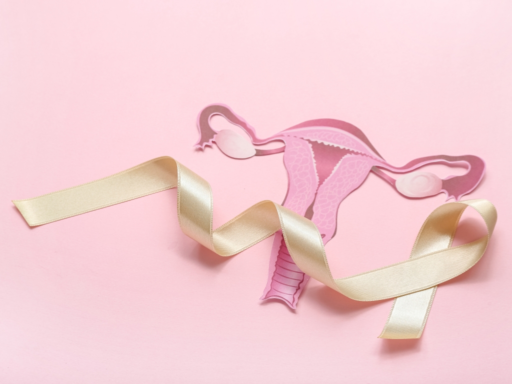
Understanding Calcified Uterine Leiomyoma
A calcified uterine leiomyoma forms when a fibroid outgrows its blood supply and begins to degenerate. During this process, calcium deposits accumulate within the tissue, making the fibroid firm or even rock-like in consistency. This transformation typically occurs later in a fibroid’s life cycle, especially in women approaching menopause.
Although calcified uterine leiomyoma is often considered inactive, it does not mean it is entirely free from hormonal influence. Hormones, particularly estrogen and progesterone, are still key factors in the earlier development stages and may continue to affect surrounding uterine tissue and symptoms.
The Role of Estrogen in Fibroid Growth
Estrogen is one of the primary hormones responsible for the growth of uterine fibroids. It stimulates the proliferation of smooth muscle cells and increases blood flow to the uterus. During reproductive years, when estrogen levels are higher, fibroids tend to grow more rapidly.
In the case of calcified uterine leiomyoma, estrogen may have played a significant role during the earlier growth phase before calcification occurred. Even after calcification, residual hormonal sensitivity in surrounding tissues may contribute to symptoms such as pelvic discomfort or pressure.
Additionally, estrogen promotes the production of growth factors that support fibroid expansion. This hormonal stimulation is why fibroids are rarely seen before puberty and often shrink after menopause when estrogen levels decline.
Progesterone and Its Influence
While estrogen is often highlighted, progesterone also plays a crucial role in fibroid development. It works synergistically with estrogen to enhance cell proliferation and inhibit apoptosis (cell death).
Research suggests that progesterone may actually have a more direct role in fibroid growth than previously believed. In calcified uterine leiomyoma, progesterone’s influence may persist in the surrounding uterine environment, even if the fibroid itself has hardened. This can contribute to ongoing symptoms such as bloating or mild pelvic pain.
Hormonal fluctuations during the menstrual cycle can also impact how a calcified uterine leiomyoma behaves, sometimes causing temporary symptom flare-ups.
Hormonal Changes During Menopause
Menopause marks a significant turning point in the hormonal landscape of a woman’s body. As estrogen and progesterone levels decline, most fibroids, including calcified uterine leiomyoma, tend to stabilize or shrink.
Calcification is more commonly observed during or after menopause because reduced blood supply and hormonal support accelerate fibroid degeneration. However, this does not mean symptoms always disappear. Some women may continue to experience discomfort due to the size or location of the calcified uterine leiomyoma.
Interestingly, hormone replacement therapy (HRT) can sometimes influence fibroid behavior. Women undergoing HRT may notice that symptoms associated with calcified uterine leiomyoma persist or slightly worsen, depending on the hormonal regimen.
Growth Factors and Hormonal Interaction
Beyond estrogen and progesterone, several growth factors and signaling pathways interact with hormones to influence fibroid development. These include transforming growth factor-beta (TGF-β), insulin-like growth factors (IGFs), and epidermal growth factors (EGFs).
In calcified uterine leiomyoma, these growth factors may have contributed to the fibroid’s initial growth and structural changes. Even after calcification, the surrounding uterine tissue may still respond to these biochemical signals, which are often regulated by hormones.
This complex interplay highlights that calcified uterine leiomyoma is not merely a static condition but part of a broader hormonal and cellular environment.
Symptoms Linked to Hormonal Influence
Although calcified uterine leiomyoma is often less symptomatic than active fibroids, hormonal influences can still trigger certain issues. Common symptoms include:
- Pelvic pressure or heaviness
- Lower abdominal discomfort
- Occasional pain during menstruation
- Urinary frequency if the fibroid presses on the bladder
Hormonal fluctuations, especially during perimenopause, can intensify these symptoms temporarily. Even though the fibroid is calcified, the uterus and surrounding tissues remain hormonally responsive.
Diagnostic Considerations
Diagnosing calcified uterine leiomyoma typically involves imaging techniques such as ultrasound, CT scans, or MRI. Calcified fibroids appear distinct due to their dense, hardened structure.
Understanding hormonal history is also crucial during diagnosis. Factors such as age, menstrual patterns, and use of hormonal medications can provide insight into how a calcified uterine leiomyoma developed and whether hormones are still influencing symptoms.
Doctors may also evaluate hormone levels to determine if additional treatment is necessary, especially in cases where symptoms persist despite calcification.
Treatment Approaches and Hormonal Management
Treatment for calcified uterine leiomyoma depends on the severity of symptoms and the patient’s overall health. In many cases, no treatment is required if the fibroid is asymptomatic. However, when symptoms are present, several options may be considered:
1. Hormonal Therapy
Hormonal medications can help regulate estrogen and progesterone levels, potentially reducing symptoms. While they may not shrink a calcified uterine leiomyoma significantly, they can improve overall uterine health.
2. Non-Surgical Procedures
Minimally invasive treatments, such as uterine artery embolization (UAE), can cut off blood supply to fibroids. Although calcified fibroids already have reduced blood flow, this approach may still be beneficial for associated symptoms.
3. Surgical Options
In cases where a calcified uterine leiomyoma causes significant discomfort or complications, surgical removal may be recommended. This could involve myomectomy or, in more severe cases, hysterectomy.
4. Lifestyle and Hormonal Balance
Maintaining a healthy lifestyle can support hormonal balance. Regular exercise, a balanced diet, and stress management may help reduce the impact of hormonal fluctuations on symptoms related to calcified uterine leiomyoma.
The Importance of Monitoring
Even though calcified uterine leiomyoma is generally considered a later-stage fibroid, regular monitoring is still important. Hormonal changes, especially during perimenopause or with hormone therapy, can influence symptoms and overall uterine health.
Routine check-ups allow healthcare providers to track any changes in size, position, or associated symptoms. This ensures timely intervention if needed and helps maintain quality of life.
Future Research and Insights
Ongoing research continues to explore the relationship between hormones and fibroid development. Scientists are investigating new therapies that target hormonal pathways more precisely, potentially offering better outcomes for women with conditions like calcified uterine leiomyoma.
Advancements in personalized medicine may also allow for tailored treatments based on an individual’s hormonal profile, improving both effectiveness and safety.
Conclusion
Hormones play a fundamental role in the lifecycle of uterine fibroids, from their initial formation to eventual calcification. While calcified uterine leiomyoma is often seen as a less active stage, hormonal influence does not completely disappear. Estrogen, progesterone, and various growth factors continue to shape the uterine environment and may contribute to ongoing symptoms.
Understanding the hormonal dynamics behind calcified uterine leiomyoma helps in making informed decisions about diagnosis, monitoring, and treatment. With proper care and awareness, women can effectively manage this condition and maintain a good quality of life, even in the presence of calcified fibroids.
When to See a Psychiatrist – A Complete Guide To Mental Health Care
Raspberry Hills Hoodie | Raspberry Hills Official Store
Affordable Hair Transplant Options Explained
Plump: The Next-Generation Crypto Casino for Modern Players
Mobile Repair in New Cumnock – Fast, Reliable & Affordable Solutions by Mobile Doctor Ayr